Fig. 11


| Fig. 10 Fig. 11 |
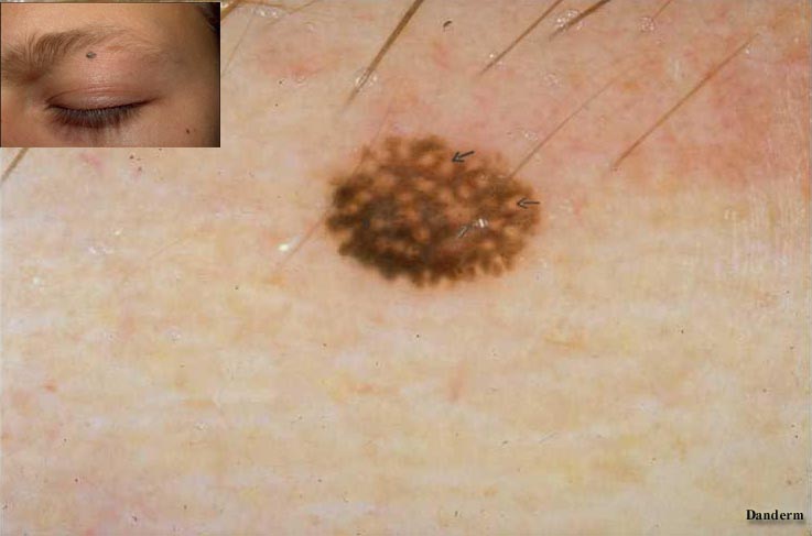
The localisation of the lesion is important, because the pigment network is different in various areas. If looking at a pigmented lesion on the face, there will be no regular network due to lack of of rete ridges. Instead there is a pseudo-pigment network due to perforations of the eccrine sweatglands (Fig.10 ®
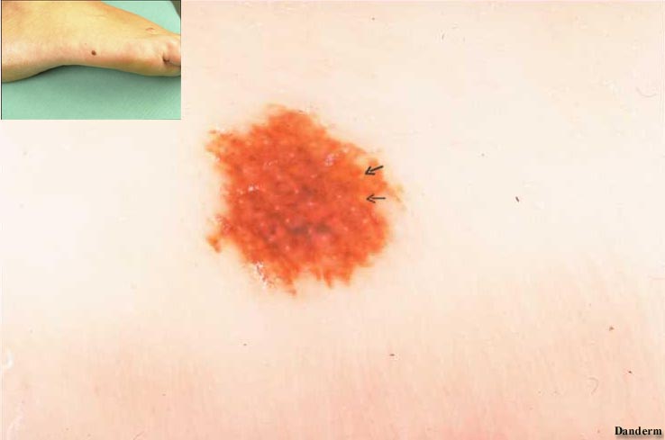
) On hands and feet the papillary rete ridges create a linear pigment pattern with openings of eccrine sweat glands appearing regularly in the middle of the less pigmented tips (Fig.11 ®
). The railway-like pigment pattern should not be mistaken for radial streaming or pseudopods of a melanoma. | |
| Fig. 12 |
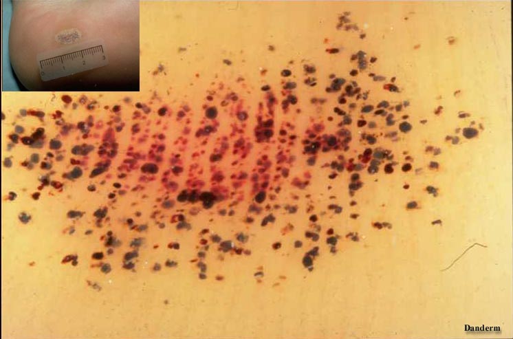
In case of mechanical trauma with cutaneous bleeding such as talon noir of e.g. badminton players, the blood pigment is primarily located in the broad tips, leaving the space between them lighter (Fig.12). |
|
|