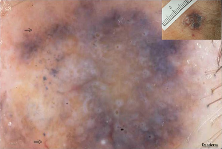
Lumps or grains of graybrownish pigmentation is typical of pigmented basalcell carcinoma (Fig.7 ® ). Often characteristic distended "retinal" vessels are observed at the periphery (Fig.7 Þ ).
Fig. 8
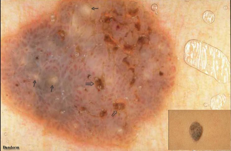
The findings are diagnostic of seborrhoeic keratosis. Horny cysts are yellowish or whitish punched-out lesions in a homogenous brownish areas without presence of a pigment network (Fig.8 ® ). Horny cysts may also occur in melanocytic naevi. Pseudofollicular openings with keratin plugging looking like comedo openings are characteristic (Fig.8 Þ ).
Fig. 9
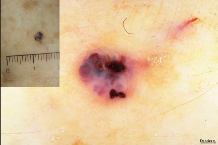
Angiomas appear as lagoons or saccular lesions filled with blood (Fig.9). There may be some crusting and darker colour due to vascular trombosis. Such lesions are clinically suspicious of malignant melanoma.

