
Fig. 21
Globules
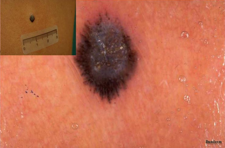
Globules are nests of pigmented melanocytes in the papillary dermis. They are defined as having a diameter of more than 0.1 mm (Fig.3® ). Spitz naevus may show a globular pattern (apart from the "frogfinger" type (Fig.21)).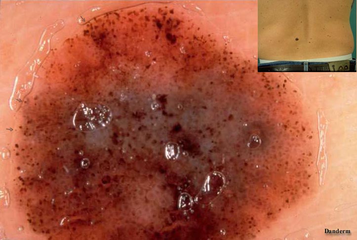
Black or darkbrown dots are less than 0.1 mm, located in the superficial layers of epidermis. Sometimes black dots are found superficially in the stratum corneum and can be removed by gentle scraping. If unevenly distributed at the periphery of a melanocytic lesion, black dots may indicate malignant melanoma as is the case of Fig.4(®)
Fig. 5
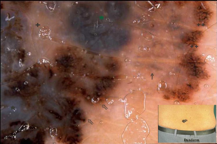
These changes are due to an abnormal pigment distribution at the junctional zone and are present in about 80% of thin malignant melanoma (< 1 mm) . Abnormal proliferating melanocytic cells distort the pigment network, producing thick dark lines at the periphery (Fig.5 ® ), sticking out at the periphery, socalled pseudopods (Fig.5 Þ ). Streaming and pseudopods are essentially identical, indicating malignant melanoma.

