Fig. 6
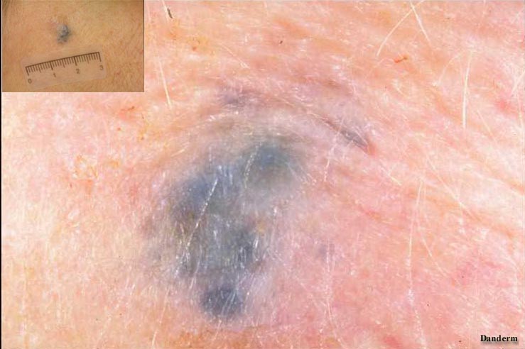
Fig. 5
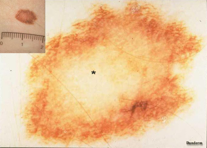
Structureless areas are seen in many melanocytic naevi due to a faint pigmentation of the keratinocytes forming the pigment network (Fig.2 * ) or in dermal naevi. Steel-blue areas are typically seen in blue naevus which is a pigmented melanocytic tumour in dermis. The pigmentation is turned bluish due to dispersion of the light (Tyndall effect) (Fig.6).
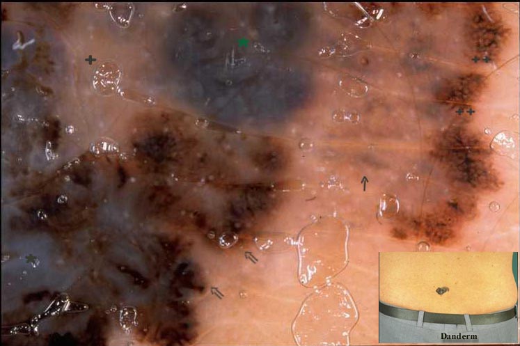
Bluegray or blackblue areas in combination with a whitish veil (due to hyperortokeratosis) (Fig.5 * ) or white scarlike areas (indicating regression) is typical of thick malignant melanoma (> 1 mm) (Fig.5 +).

