DERMATOSCOPY


Fig. 1
Click to view full size


A dermatoscope is a hand-held skin surface microscope with approximately 10 x magnification (Fig.1). The skin surface is illuminated by a halogen bulb powered with batteries or a rechargeable system built in the handle. By using immersion oil or a disinfectant liquid and by pressing the glass disc of the scope on the skin, details of epidermis and the junction zone become visible. Hereby it is possible to study the pigment pattern of the lesions, especially the pigment network which is the main object of dermatoscopic investigation.


Fig. 2
Click to view full size
Fig. 3
Click to view full size
The pigment network

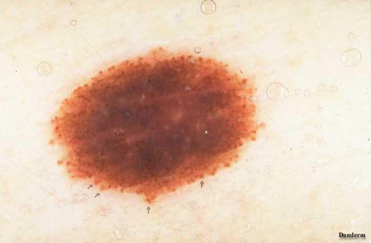
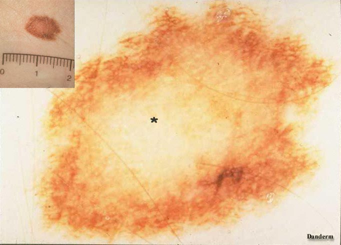
The network is formed by presence of melanin in the basal keratinocytes of the epidermal rete ridges (Fig.2). Most melanocytic naevi and melanomas have a pigment net, but abscence does not exclude a melanocytic lesion. Especially congenital melanocytic naevi have a globular pigment pattern (Fig.3). Furthermore, malignant melanoma may end up with a completely distorted or destroyed pigment net due to melanocytic proliferation (see later).